Challenges. Solutions. Results.
Successfully delivered solutions are the perfect showcase for the technical depth and breadth of our Biorep teams and the quality-is-first approach we apply to every single project.
Successfully delivered solutions are the perfect showcase for the technical depth and breadth of our Biorep teams and the quality-is-first approach we apply to every single project.
Note: Due to the confidential nature of the work we do, the following is only a sampling of our projects.
Challenge:
A next generation instrument that would deliver knots tied in the extracorporeal space, and delivered to an intracorporeal target site in a limited access setting. Device needed to have secure suture retention characteristics, eliminate the hand fatigue (normally caused by existing mechanized knot delivery devices), and needed to be manipulatable in fields where access and space is extremely limited.
Solution:
Designed a device in which the primary position of the suture eyelet gate is closed (as opposed to open) in order to address hand fatigue; the moving eyelet gate closes fully to complete circumferential suture entrapment (to address suture retention security); and with an extremely low profile and offset tip to address limited access/space settings.
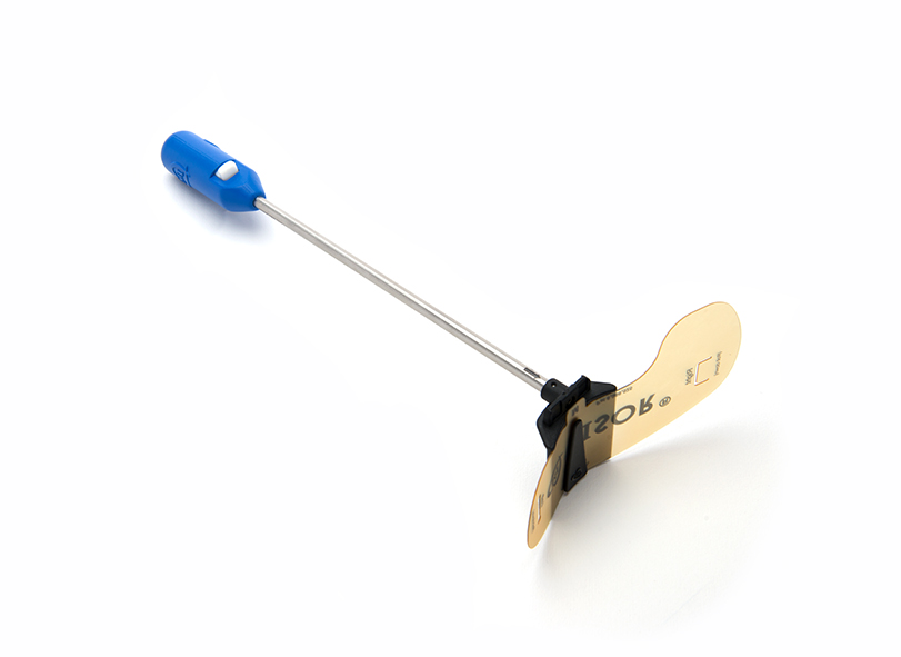
Challenge:
Need for a next gen retraction instrument that would retract the atrial wall in a circumferential manner in a limited access setting. Device needed to have: different sizes to accommodate varying atrium dimensions; be easily and securely assembled/disassembled in fields where access and space is extremely limited; and facilitate delivery of CO2 into the working space.
Solution:
Designed a device with: multiple component sizes to address varying atrium dimensions; with a flexible component (VISOR) which could be rolled into a smaller dimension and “slide” into a corresponding locking-support structure (S-BLADE) easily/efficiently/securely; and has a flexible component with the ability to “unfurl” under its own biasing to provide circumferential atrial wall retraction. The stabilization component (S-POST) which attaches to the support structure utilizes a “ball and socket” style engagement mechanism for ease of assembly/disassembly, has a CO2 port, and an open inner lumen in which CO2 can be delivered to target site.
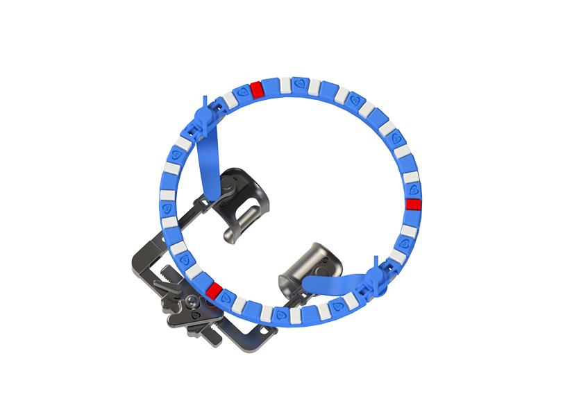
Challenge:
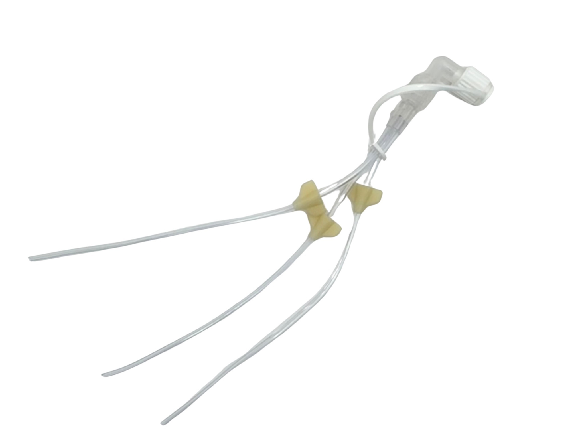
Need for a next gen “rib spreader” that would retract the intercostal space and facilitate retraction of the ribs and corresponding rib deflection movement in a bilaterally equal manner. Device needed to have accompanying retractor blades that could also contour to extreme anatomical curvatures to eliminate localized pressure points on individual ribs. Needed an accessory attachment that would accommodate simple, secure and efficient suture organization to eliminate clutter from the field, and also provide additional retraction to target site structures.
Solution:
Designed a retractor rack with arms that move outwardly and inwardly in a bilaterally equal manner when the turnkey knob is rotated. Designed set of accompanying retractor blades with separated tissue engagement surfaces that swivel independently from each other in order to contour with extreme curvatures and equally distribute pressure across the entire engagement surface of individual ribs. The design incorporates a low-profile disposable accessory component that can attach directly onto the top of the retractor arms, and is circumferential to accommodate simple and efficient organization. The design utilizes flexible wedge plugs for secure and efficient suture capture and release, and can provide additional retraction to target site structures by anchoring retraction sutures at any point around the circumference.
Challenge:
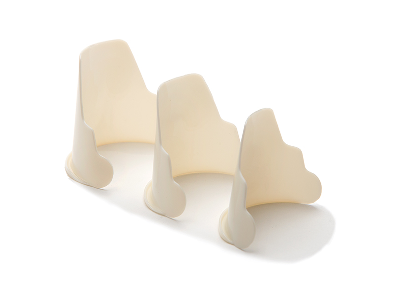
Need for a “self-sustaining” retraction instrument that would provide circumferential retraction to the aortic walls without the need for additional operator manipulation in a limited access setting. Device needed to be easily delivered and positioned and then retract the aortic wall safely and securely without the need for supportive assistance in order to allow the operator to use both hands, and without having the need for additional support instruments/structures in the working field. Need for different sizes to accommodate varying aortic root dimensions.
Solution:
Designed a flexible retractor which rolls into a smaller dimension and deliverable into aortic root easily, efficiently, and securely. The flexible retractor can “unfurl” under its own biasing to provide circumferential atrial wall retraction. Designed with a flange on the bottom of the device which would engage with the sino-tubular junction in order to keep the device securely seated in-situ without the need for additional manipulation. Design uses a soft and smooth material with all outer edges curved, blunted and atraumatic for safe delivery and deployment purposes, and incorporated this design into three different sizes to accommodate varying aortic root dimensions.
Challenge:
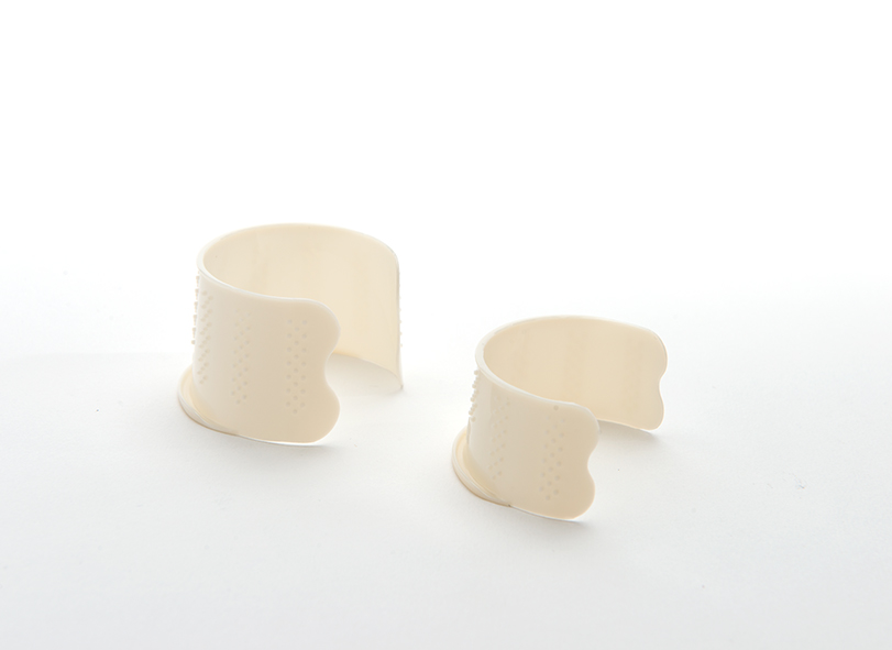
Need for a “self-sustaining” instrument that would provide circumferential retraction to the mitral valve leaflets in order to enable access and exposure to the intra-ventricular site and the sub-valvular apparatus without the need for additional operator manipulation in a limited access setting.
Device needed to be able to be easily delivered and positioned, then able to retract the mitral valve leaflets safely and securely without the need for supportive assistance in order to allow the operator to use both hands without having the need for additional support instruments or structures in the working field. Need for different sizes to accommodate varying mitral valve annular dimensions.
Solution:
Designed a flexible retractor which could be rolled into a smaller dimension and delivered into the mitral annulus easily/efficiently/securely and then the flexible retractor has the ability to “unfurl” under its own biasing to provide circumferential mitral valve leaflet retraction. Designed a flange on the leading edge of the device which would engage with the posterior side of the mitral annulus to prevent the device from “popping out”, and added traction raisings on the outer device wall surface to engage with the mitral valve leaflets to prevent the device from rotating in order to keep the device securely seated in-situ without the need for additional manipulation. Design uses a soft and smooth material with all outer edges curved, blunted and atraumatic, for safe delivery and deployment purposes, and incorporated design into two different sizes to accommodate varying mitral valve annular dimensions.

Challenge:
As cardiac surgery continues to shift toward minimally invasive and robotic approaches, surgeons must operate through smaller incisions with reduced access to the ascending aorta. This makes achieving safe, controlled, and reproducible cross-clamping more complex, often introducing variability in placement and adding inefficiencies to procedural workflow.
Solution:
An advanced aortic cross-clamp system was developed to enable precise and controlled occlusion across both open and minimally invasive procedures. The system incorporates a detachable clamp head and a dedicated delivery mechanism, allowing surgeons to position and secure the clamp with confidence before removing the handle to maintain a clear operative field. Its design supports reliable vessel occlusion with controlled force application, stable positioning without obstructing access, and improved maneuverability in constrained environments, while integrating seamlessly into minimally invasive and robotic workflows.

Challenge:
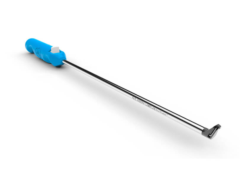
Minimally invasive cardiac procedures require surgeons to perform precise suture knot placement within confined operative spaces and limited visualization. Traditional techniques can be difficult to control in these environments, increasing procedural complexity and variability, particularly during critical steps such as neochord placement.
Solution:
An intracorporeal knot placement assist device was developed to enable accurate and controlled suture knot positioning in minimally invasive cardiac procedures. The device incorporates a low-profile tip designed for precise placement at the target site, along with a suture loop mechanism that enhances grip and tension control during knot delivery. Its ergonomic design supports consistent handling and reduces the effort required to maintain stable positioning within confined spaces, helping surgeons achieve reliable knot placement with improved control and efficiency.
Circulogix, Inc., a Miami, Florida based medical device company for clinical cancer research, partnered with Biorep Technologies as its manufacturing partner.
The company commercializes the faCTChecker™ — a fully automated, size-based circulating tumor cell (CTC) capture instrument — and the CyteCatch™ microfilter slide, the core consumable enabling blood-based cancer cell isolation for clinical research, biomarker discovery, and longitudinal clinical trials.
Biorep’s manufacturing precision and quality systems have enabled Circulogix to supply systems to some of the most rigorous cancer research environments in the United States.

The irrigation syringe attachment represents a breakthrough in treating Hidradenitis Suppurativa tunneling wounds (HS TW), offering a minimally scarring and non-invasive method that improves patient tolerance and simplicity.
© 2026 Biorep Technologies, Inc. All rights reserved.